March 2020
Fetal Cardiac Intervention
By Stephanie Guseh, MD, Fetal Therapy SIG
Congenital heart disease (CHD) affects approximately 1% of all births every year. Of children born with CHD, 25% of them are considered severe and will require surgery or other invasive procedures within the first year of life. As prenatal imaging and diagnosis has improved, so too has our understanding of the natural history of many of these congenital malformations. In some cases, specific cardiac lesions have been shown to predictably evolve throughout pregnancy from causing mild-to-moderate impairment to more severe, even life-limiting disease. As with all types of fetal intervention, it is this subset of particularly severe cases with an opportunity for mid-gestation intervention, that are the focus of fetal cardiac intervention (FCI).
Initial attempts at FCI were first reported in the early 1990s but were limited by high rates of morbidity and mortality. Over the next two decades, significant improvements in prenatal imaging, anesthesia, balloon catheters, and our understanding of the natural history of CHD prompted several multi-disciplinary teams to re-evaluate the approach to FCI. The importance of this multi-disciplinary collaboration cannot be overstated, as the teams comprise representatives from fetal cardiology, interventional cardiology, maternal fetal medicine, fetal anesthesiology, maternal anesthesiology, nursing, and social work, among others. These disciplines work together to identify the cardiac lesions most amenable to fetal intervention and to minimize the associated maternal and fetal risks. To date, the focus of FCI has been primarily directed on three lesions, namely severe aortic stenosis (AS) with evolving hypoplastic left heart syndrome (eHLHS), HLHS with a highly restrictive or intact atrial septum, and pulmonary atresia with an intact ventricular septum (or hypoplastic right heart syndrome). In each of these CHDs, the goal of intervention is to improve post-natal outcomes either by salvaging some cardiac function and retaining the possibility of biventricular circulation after birth, by minimizing collateral damage to the lungs and vasculatures, or by improving the immediate stabilization after birth.
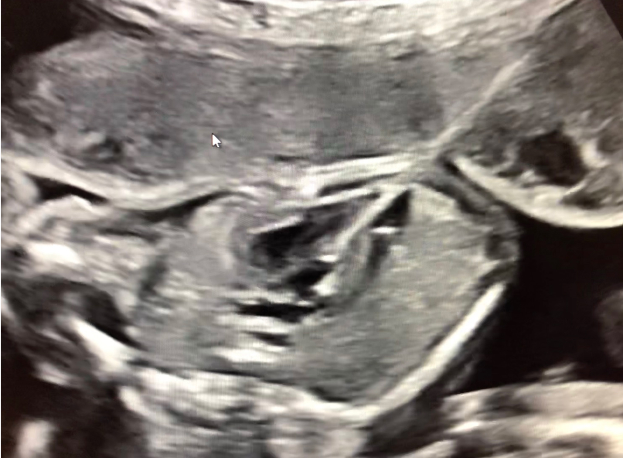
Although initial data on our ability to achieve the above outcomes seem promising, the goals of these interventions must be balanced against the maternal and fetal risks associated with each procedure. (Gardiner, Ultrasound Obstet Gynecol 2016; Friedman, Ultrasound Obstet Gynecol 2018; Freud, Circulation 2014) Improvements in technique over the last 20 years have led to a majority of these procedures now being performed under regional anesthesia with a minimally-invasive 18- or 19-guage cannula, as seen in Figure 1. These procedural improvements along with more refined fetal resuscitation have improved rates of fetal loss, which generally lies between 5-10%. (Mizrahi-Arnaud, Pediatr Res 2007) Minimizing the risk of fetal loss is one important component of the continual process of improvement that must accompany these invasive procedures. In addition to improving both maternal and fetal safety associated with FCI, ongoing research efforts are aimed at optimizing candidate selection, identifying accurate predictors of success through genetic testing and biomarker availability, and evaluating the long-term cardiac and neurodevelopmental outcomes of these procedures. (Friedman, Prenat Diagn 2018; Laraja, J Pediatr 2017)
Despite the improvement in technical success and the exciting research advances detailed above, many controversies surrounding FCI still exist. Through collaboration and discussion facilitated by the International Society of Prenatal Diagnosis, the Fetal Therapy SIG aims to both support the advances of and enable a discussion of the challenges surrounding FCI. Through international and multi-disciplinary collaboration, we hope to provide both information for patients as well as a forum for professional discourse regarding the role of FCI in improving outcomes for those pregnancies diagnosed with severe CHD.
References
Freud LR, McElhinney DB, Marshall AC, et al. Fetal aortic valvuloplasty for evolving hypoplastic left heart syndrome: postnatal outcomes of the first 100 patients. Circulation. 2014 Aug 19;130(8):638-45. doi: 10.1161/CIRCULATIONAHA.114.009032. Epub 2014 Jul 22. PubMed PMID: 25052401; PubMed Central PMCID: PMC4299861.
Friedman KG, Sleeper LA, Fichorova RN, et al. Myocardial injury in fetal aortic stenosis: Insights from amniotic fluid analysis. Prenat Diagn. 2018 Feb;38(3):190-195. doi: 10.1002/pd.5213. Epub 2018 Feb 6. PubMed PMID: 29327361.
Friedman KG, Sleeper LA, Freud LR, et al. Improved technical success, postnatal outcome and refined predictors of outcome for fetal aortic valvuloplasty. Ultrasound Obstet Gynecol. 2018 Aug;52(2):212-220. doi: 10.1002/uog.17530. Epub 2018 Jul 4. PubMed PMID: 28543953.
Gardiner HM, Kovacevic A, Tulzer G, et al.; Fetal Working Group of the AEPC. Natural history of 107 cases of fetal aortic stenosis from a European multicenter retrospective study. Ultrasound Obstet Gynecol. 2016 Sep;48(3):373-81. doi: 10.1002/uog.15876. Epub 2016 Aug 4. PubMed PMID: 26843026.
Laraja K, Sadhwani A, Tworetzky W, et al. Neurodevelopmental Outcome in Children after Fetal Cardiac Intervention for Aortic Stenosis with Evolving Hypoplastic Left Heart Syndrome. J Pediatr. 2017 May;184:130-136.e4. doi: 10.1016/j.jpeds.2017.01.034. Epub 2017 Feb 21. PubMed PMID: 28233547; PubMed Central PMCID: PMC6343658.
Mizrahi-Arnaud A, Tworetzky W, Bulich LA, et al. Pathophysiology, management, and outcomes of fetal hemodynamic instability during prenatal cardiac intervention. Pediatr Res. 2007 Sep;62(3):325-30. PubMed PMID: 17622948.
Figure 1. Intra-procedural image of a minimally-invasive fetal aortic valvuloplasty for aortic stenosis with evolving hypoplastic left heart syndrome.